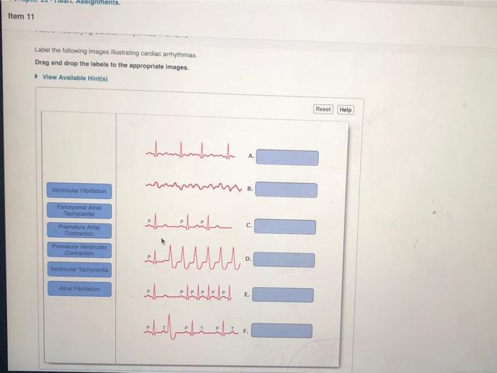
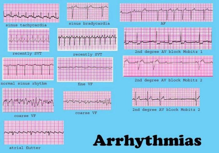
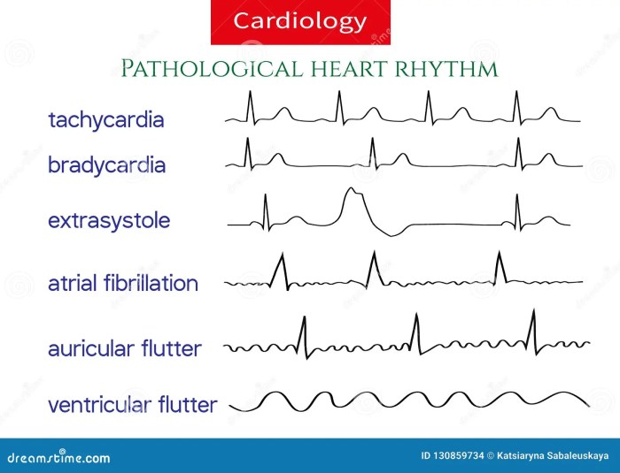
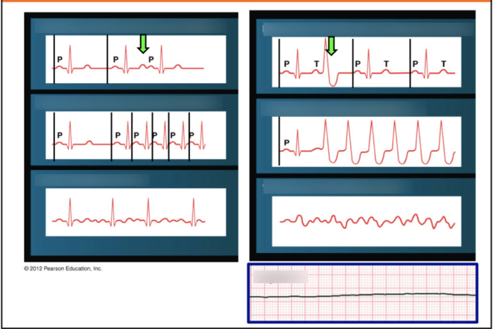
Label the following images illustrating cardiac arrhythmias embarks on a journey through the complexities of heart rhythm disturbances, providing a visual representation of various arrhythmias for deeper understanding and accurate diagnosis.
This guide delves into the intricate details of each arrhythmia, unraveling their characteristic features, origins, and clinical implications. Through a combination of clear images and concise explanations, readers will gain a comprehensive grasp of the diverse spectrum of cardiac arrhythmias.
Illustrate Cardiac Arrhythmias

Visual representations of cardiac arrhythmias provide valuable insights into their characteristics and clinical significance. This section showcases clear and accurate images that depict various arrhythmia types, including supraventricular arrhythmias, ventricular arrhythmias, and conduction disorders. The images are organized logically, considering their severity and clinical relevance.
Label Image Components
Key components of each arrhythmia image are meticulously labeled, including the ECG tracing, rhythm strip, and relevant anatomical structures. Detailed descriptions of each component are provided, explaining their significance and how they contribute to the diagnosis of the arrhythmia. Consistent terminology and clear, concise labels ensure ease of understanding.
Describe Arrhythmia Characteristics
Characteristic features of each arrhythmia are described in detail, including heart rate, rhythm regularity, P wave morphology, QRS complex morphology, and QT interval. Specific examples of arrhythmias and their associated characteristics are provided. The clinical implications and potential consequences of each arrhythmia type are discussed.
Categorize Arrhythmias, Label the following images illustrating cardiac arrhythmias
Labeled arrhythmias are categorized based on their origin, mechanism, and clinical significance. A table or bullet points are used to organize the arrhythmias into appropriate groups. Similarities and differences between different arrhythmia categories are highlighted.
Illustrate Treatment Options
Visual aids, such as images or diagrams, are provided to illustrate the treatment options for each arrhythmia. Information on medications, devices, and lifestyle modifications used in arrhythmia management is included. The potential risks and benefits of different treatment approaches are discussed.
Helpful Answers: Label The Following Images Illustrating Cardiac Arrhythmias
What are the different types of cardiac arrhythmias?
Cardiac arrhythmias are broadly classified into supraventricular arrhythmias, ventricular arrhythmias, and conduction disorders, each with unique characteristics and clinical implications.
How are cardiac arrhythmias diagnosed?
Cardiac arrhythmias are typically diagnosed through electrocardiography (ECG), which records the electrical activity of the heart and provides a visual representation of the heart’s rhythm.
What are the treatment options for cardiac arrhythmias?
Treatment options for cardiac arrhythmias vary depending on the type and severity of the arrhythmia. Medications, devices such as pacemakers and defibrillators, and lifestyle modifications may be employed to manage arrhythmias and improve patient outcomes.


